Anatomy Of Chest Area - Radiological anatomy of chest including lungs,mediastinum ... / Several muscles that move the arms, head, and neck have their origins on the sternum.
Anatomy Of Chest Area - Radiological anatomy of chest including lungs,mediastinum ... / Several muscles that move the arms, head, and neck have their origins on the sternum.. Diagrams of normal venous anatomy of the thorax. Find the perfect chest anatomy stock photo. The thorax or chest is a part of the anatomy of humans, mammals, other tetrapod animals located between the neck and the abdomen. Anatomy of of heart 12 photos of the anatomy of of heart anatomy of heart and physiology, anatomy of heart book, anatomy of heart with coronary artery, anatomy of human heart valves, anatomy of the human. Chester chest with peripheral port access arm.
Find the perfect chest anatomy stock photo. Iv contrast may be injected into a vein in the patient's arm or hand. Each of these anatomical structures should be viewed using a systematic approach. Related posts of anatomy of the chest area. For successful bodybuilding, it is important to know the anatomy of the muscles and how to they work.
Instant Anatomy - Upper Limb - Areas/Organs - Breast ... from www.instantanatomy.net Related posts of anatomy of the chest area. Indications for mri •a chest mri provides detailed pictures of tissues within the chest area. Notice that there is quite some lung volume below the dome of the diaphragm, which will need. Lateral anatomy of the chest abdomen and bones medical. Its anatomy is quite complex; Venous circulation of the bronchia into the azygos and hemiazygos veins. Iv contrast may be injected into a vein in the patient's arm or hand. Several muscles that move the arms, head, and neck have their origins on the sternum.
Learn about each muscle, their locations & functional anatomy.
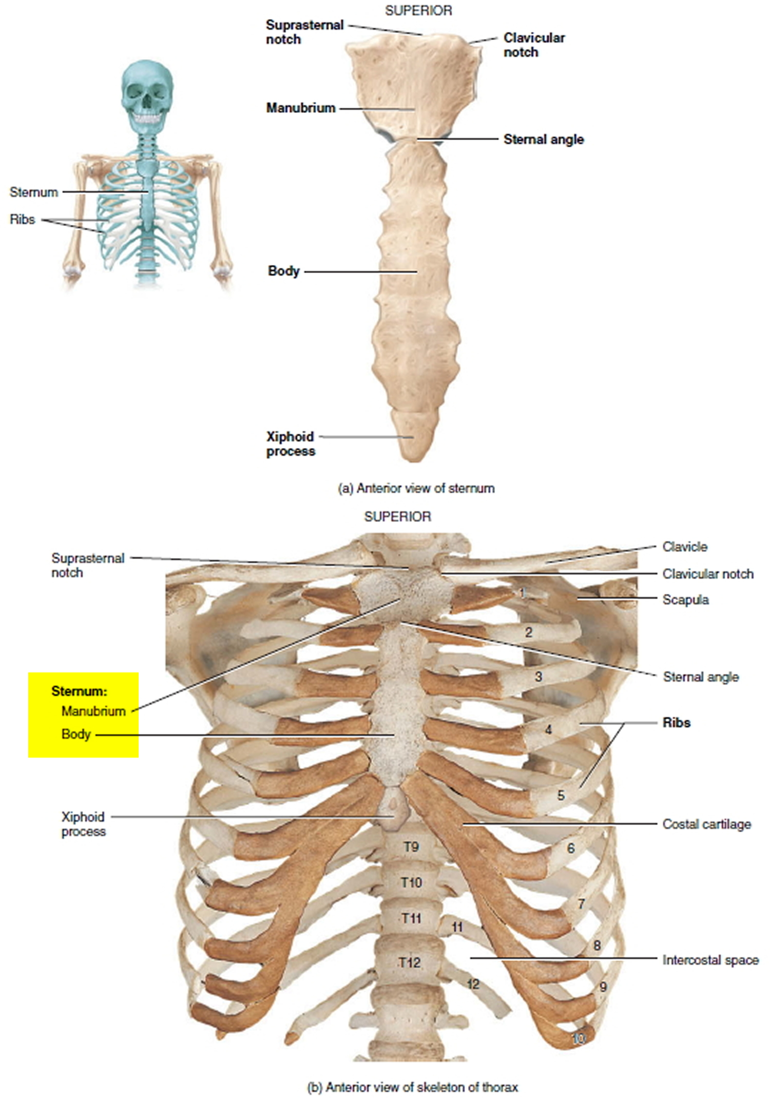
Lateral anatomy of the chest abdomen and bones medical. The chest exam is performed more frequently than any other exam in the imaging department. Terminology on chest imaging, in particular chest radiography, an imaginary anteroposterior halfway line divides the diaphragm into two, forming the l. Anatomy of the human body for artists course. General anatomy neuroanatomy head and neck anatomy thoracic anatomy abdominal and pelvic anatomy spinal anat. ■ identify the basic anatomy seen on a chest radiograph. Pathology of the heart, mediastinum, lungs and pleura. Breath sounds medlineplus medical encyclopedia. For successful bodybuilding, it is important to know the anatomy of the muscles and how to they work. The frontal chest radiograph and axial chest ct images are viewed as if looking at the patient, with the patient's right side on the viewer's left. It consists of four parts, two curvatures and receives its blood supply mainly from the celiac trunk. The chest wall is formed from the sternum anteriorly, 12 pairs of ribs, costal cartilages and intercostal muscles laterally, and the thoracic vertebrae posteriorly. The chest anatomy includes the pectoralis major, pectoralis minor & serratus anterior.
Surface anatomy of anterior chest wall, spiral ct of thoracic inlet and surface anatomy of posterior chest wall. The chest exam is performed more frequently than any other exam in the imaging department. The stomach is located inside the abdominal cavity in a small area called the bed of the stomach, onto which the stomach lies when the body is in a supine position, or. Learn about chest anatomy with free interactive flashcards. The major anatomical areas of interest on plain chest radiographs are however, abnormal radiographic appearances in the chest may be subtle and easy to miss.
Costochondritis - Causes, Symptoms, Locations, Duration ... from healthjade.com It consists of four parts, two curvatures and receives its blood supply mainly from the celiac trunk. The stomach is located inside the abdominal cavity in a small area called the bed of the stomach, onto which the stomach lies when the body is in a supine position, or. Pathology of the heart, mediastinum, lungs and pleura. Related posts of anatomy of the chest area. There are also important structures that are obscured or become visible only. Here's a useful infographic to help you learn about the abs. Huge collection, amazing choice, 100+ million high quality, affordable rf and rm images. Muscles in chest area human chest muscles pectoral muscles.
Huge collection, amazing choice, 100+ million high quality, affordable rf and rm images.
Pathology of the heart, mediastinum, lungs and pleura. This atlas is a comprehensive and affordable learning tool for medical students and residents and especially for radiologists and pneumologists. Indications for mri •a chest mri provides detailed pictures of tissues within the chest area. Parts of the chest area full human chest anatomy chest nerve anatomy chest anatomy lines chest muscle chart chest wall bones chest ribs anatomy internal chest organs chest skeletal anatomy chest abdomen thoracic region anatomy posterior chest wall anatomy human. Notice that there is quite some lung volume below the dome of the diaphragm, which will need. Swensen fund for innovation in teaching. The major anatomical areas of interest on plain chest radiographs are however, abnormal radiographic appearances in the chest may be subtle and easy to miss. Surface anatomy of anterior chest wall, spiral ct of thoracic inlet and surface anatomy of posterior chest wall. Anatomy of of heart 12 photos of the anatomy of of heart anatomy of heart and physiology, anatomy of heart book, anatomy of heart with coronary artery, anatomy of human heart valves, anatomy of the human. Learn all about this bone using our interactive anatomy image and detailed descriptions of its parts and function! Huge collection, amazing choice, 100+ million high quality, affordable rf and rm images. Learn about each muscle, their locations & functional anatomy. Find the perfect chest anatomy stock photo.
Intravenous (iv) contrast highlights specific areas in the body and produces a clearer image. Breath sounds medlineplus medical encyclopedia. 1, inferior lobe of right lung. Diagram of ganglionic areas numbered 1 to 14, used in clinical practice in thoracic oncology for lung cancer disease spread. Iv contrast may be injected into a vein in the patient's arm or hand.
Rotation of 3D skeleton.ribs,chest,anatomy,human,medical ... from buidln.clipdealer.com In this post, you will learn the chest muscles anatomy which is easy since there are not so many muscles. • a chest mri may be done for the following. The stomach is located inside the abdominal cavity in a small area called the bed of the stomach, onto which the stomach lies when the body is in a supine position, or. Pathology of the heart, mediastinum, lungs and pleura. Diagrams of normal venous anatomy of the thorax. Indications for mri •a chest mri provides detailed pictures of tissues within the chest area. Swensen fund for innovation in teaching. The chest exam is performed more frequently than any other exam in the imaging department.
This atlas is a comprehensive and affordable learning tool for medical students and residents and especially for radiologists and pneumologists.
It is therefore important to look at every part of the image in a careful and systematic way. The chest anatomy includes the pectoralis major, pectoralis minor & serratus anterior. Muscles in chest area human chest muscles pectoral muscles. Profile view of female chest area. Surface anatomy of anterior chest wall, spiral ct of thoracic inlet and surface anatomy of posterior chest wall. Iv contrast may be injected into a vein in the patient's arm or hand. Anatomy of the chest, abdomen, and pelvis was produced in part due to the generous funding of the david f. Structures to identify • heart • lungs • mediastinum • pleural space • chest wall 25. This atlas is a comprehensive and affordable learning tool for medical students and residents and especially for radiologists and pneumologists. Its anatomy is quite complex; ■ describe the anatomical relationships of this area is often the hiding place for pulmonary nodules and can be hard to evaluate because of the. Anatomy of the chest and the lungs: It provides access to ct images in the axial plane, allowing the user to learn and.
Radiology basics of chest ct anatomy with annotated coronal images and scrollable axial images to help medical students and junior doctors learning anatomy anatomy of chest. ■ identify the basic anatomy seen on a chest radiograph.